 Nix McRetro
After having a headache for nearly a week, I figured I should get it checked out at the local GP. Thanks to having a somewhat decent health system in place, I had a CT scan just one hour later. Having a background in human biomedical science, I naturally started poking around the images myself. Nothing obvious jumped out at me, apart from what looked like a chunky calcified or ossified section of the falx cerebri. The radiology report did not identify it as pathology, so this was me admiring an incidental-looking bit of anatomy rather than diagnosing myself.
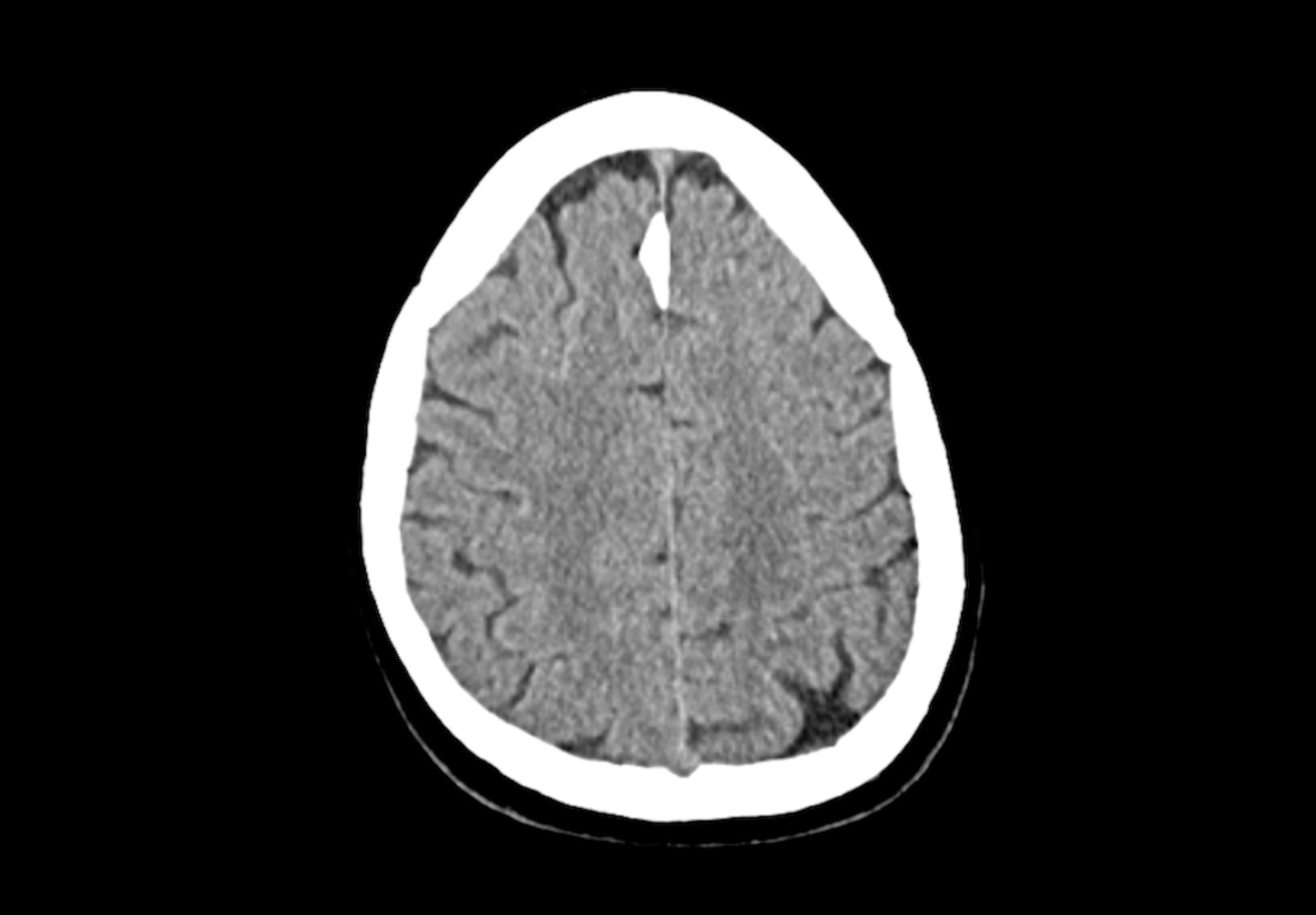
The CT scans provided have two display settings: brain window and bone window. Above is the brain window, which shows more detail in the brain itself. The falx cerebri is visible as that bright white section.
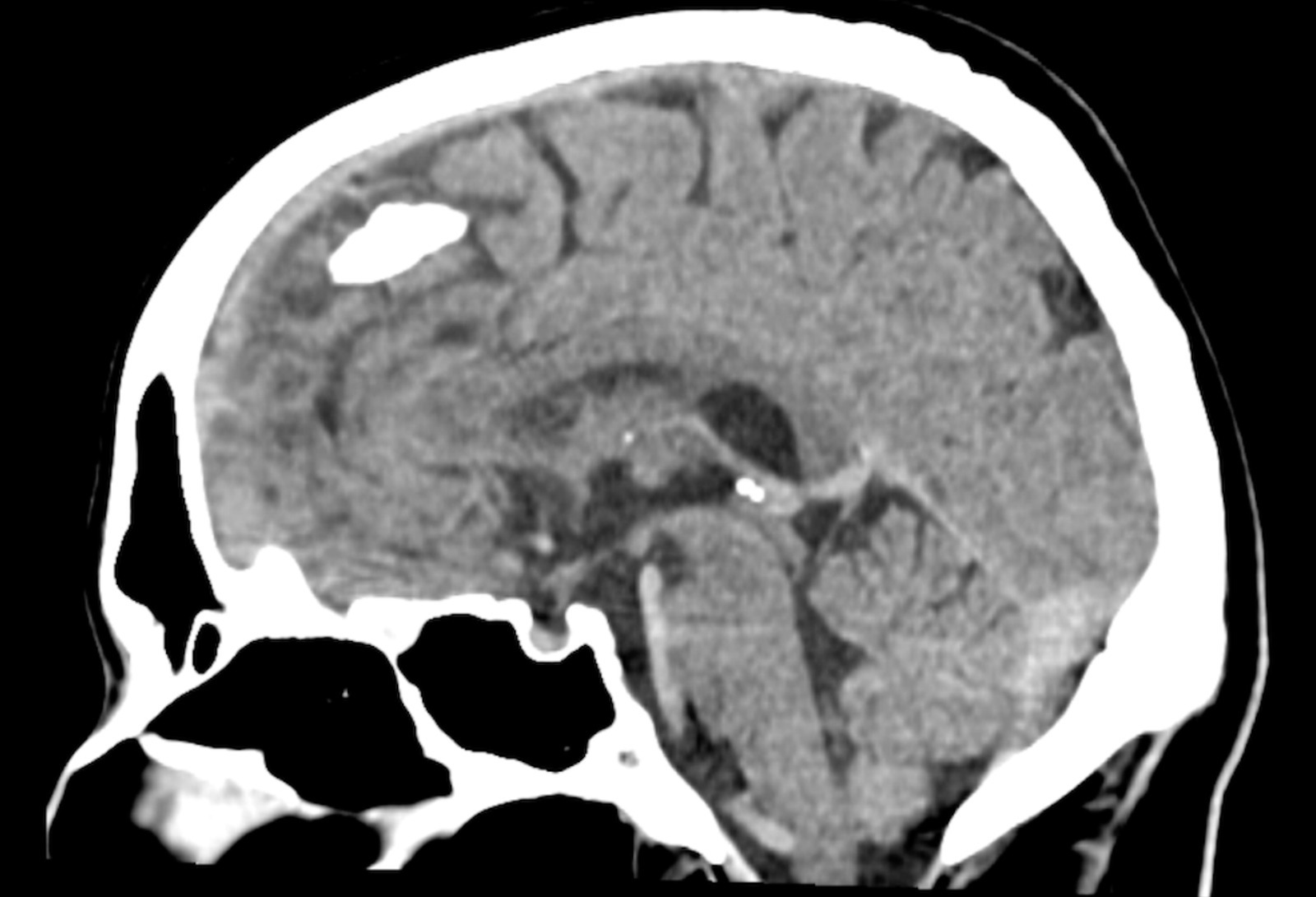
After a quick anatomy refresher, the falx cerebri is the dural fold separating the left and right cerebral hemispheres. Calcification and true ossification are not quite the same thing, and I couldn’t establish which I was looking at from these screenshots alone. Both falx calcification and ossification can turn up as incidental imaging findings.
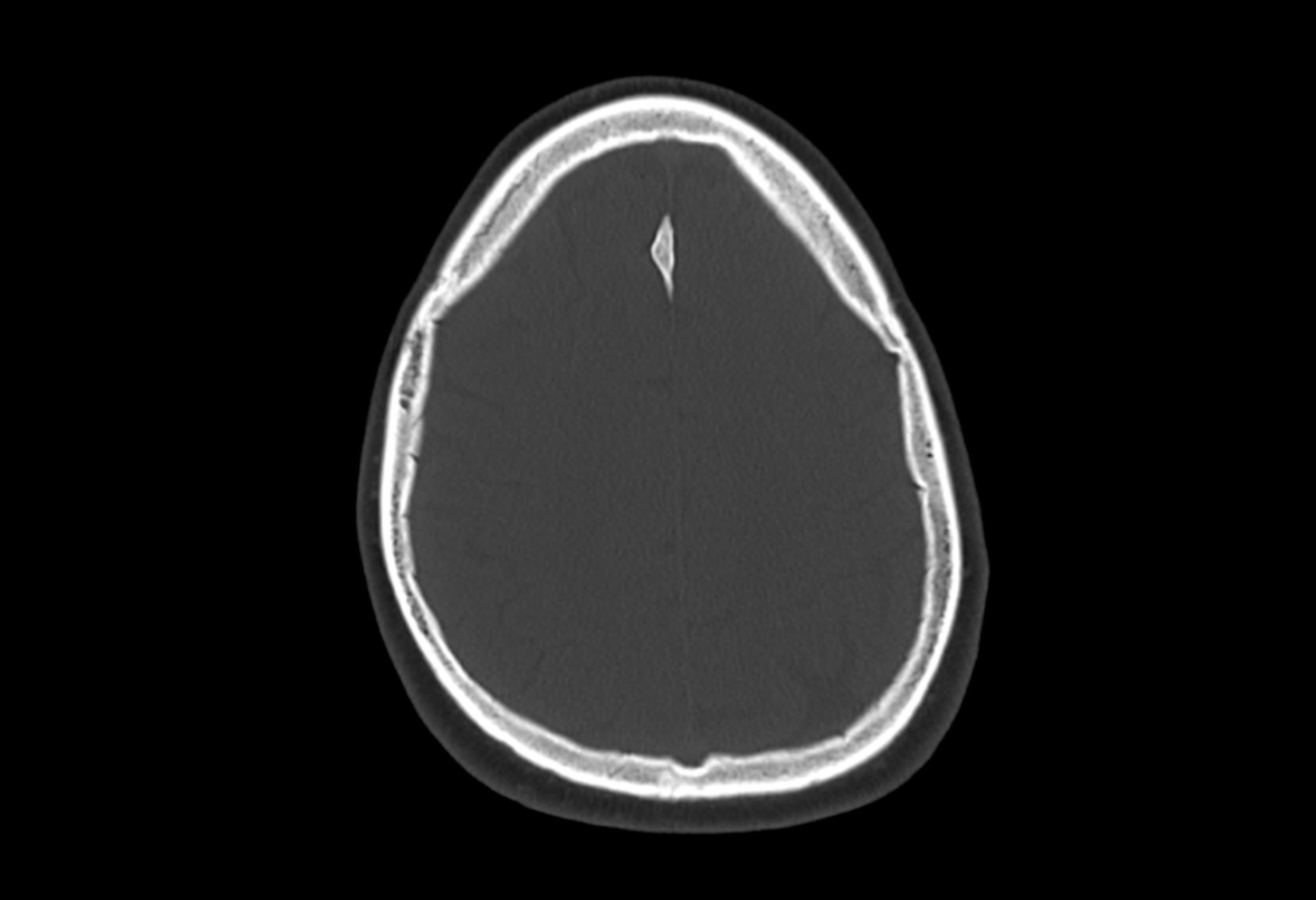
Next we have bone window mode, where you can see the bone detail but not so much the brain squiggly mush bits, I want to say spaghetti? To my eyes, the bright section of falx looked very similar to the skull itself. That alone doesn’t establish whether it was calcification or true ossification, but it certainly looks neat. Cool science!
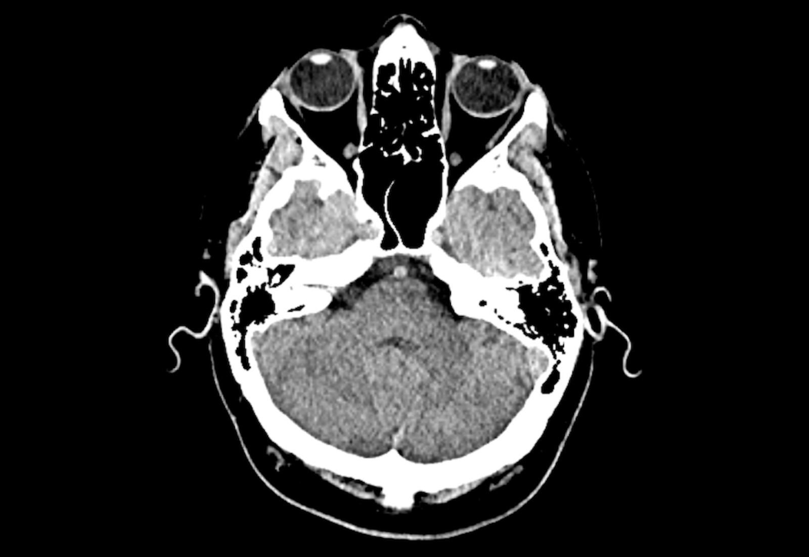
Unfortunately, none of this helps my headache. To make matters worse, this headache doesn’t seem to be affected by ibuprofen, naproxen or paracetamol. I need to be doing other things in this down time but I can’t think straight for long before my head starts to hurt. Is it stress? Probably. My eye twitch disappeared just before this started… last Wednesday. 😅
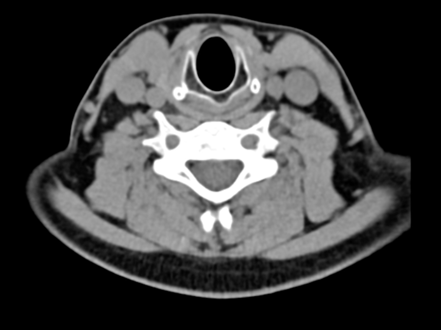
I love pattern recognition though, is that a happy axolotl hiding in my vertebrae? A little less spooky than the other image with the eyeballs. Here’s the full report from the doctor.
Technique:
Non-contrast study of the head and cervical spine was performed and the images were reviewed in multiple planes.
Findings:
Brain
There is normal attenuation of the brain with normal grey-white matter interface. There is no acute intracranial bleed or established acute infarct. Ventricles are non-dilated. There is incidental finding of CVI (cavum velum interpositum- anatomic variation), that measures up to 18 mm transverse the medial and 15 mm craniocaudal with convex margins indicating some degree of mass effect. Consider further imaging with MRI. There is no brain oedema, or midline shift. The rest of the CSF spaces define normally. The bone window images of the skull do not reveal any abnormality.
Cervical Spine
There is normal alignment of the cervical vertebrae. The facet joints show normal articulation. The odontoid is intact and normal in position. The CV junction and atlantoaxial articulation are normal. There is congenital nonfusion of the posterior arch of C1. There is no significant disc osteophyte complex or spinal canal stenosis. There is no neural foraminal narrowing. The cervicothoracic junction is in normal alignment. There is no prevertebral or paraspinal collection.
Conclusion:
No significant intracranial pathology. Incidental finding of anatomic variation that may need further characterisation with MRI to assess for any CSF flow abnormality. No significant cervical spine pathology.
Interestingly, the falx was not the finding the radiologist actually wanted followed up. That was the cavum velum interpositum (CVI). A CVI is a CSF-filled midline anatomical variant and is generally considered part of the normal spectrum, although enlargement or a cyst producing mass effect can warrant further investigation. In my scan the radiologist specifically noted convex margins and some degree of mass effect, which is why the MRI was recommended.
Sources
- Radiology Masterclass - Intracranial calcification
- Radiopaedia - Falx ossification
- Falx Cerebri Ossification: CT and MRI Evaluation
- Imaging of the septum pellucidum: normal, variants and pathology